Credits
Cost
Reviews
Why RadComm
Credits
 This self-directed course is worth 8 Category A continuing education credits. RadComm Courses are Approved by the AHRA and Recognized by the ARRT.
Cost
 The Course Costs $97.00 USD for a full year of access. All RadComm courses are backed by a full money-back guarantee.
Reviews
[woocommerce_reviews_avgstars]
[woocommerce_reviews_avgtext]
[woocommerce_reviews_number]
Why RadComm
RadComm courses are designed for busy techs just like you. Each self guided course is:
- Approved: by an MQSA RCEEM recognized by the ARRT & all 50 states.
- On-Demand: Immediate access after enrollment; Study at your own pace.
- Informative: Designed by leading breast educators to improve your patient care & breast health knowledge.

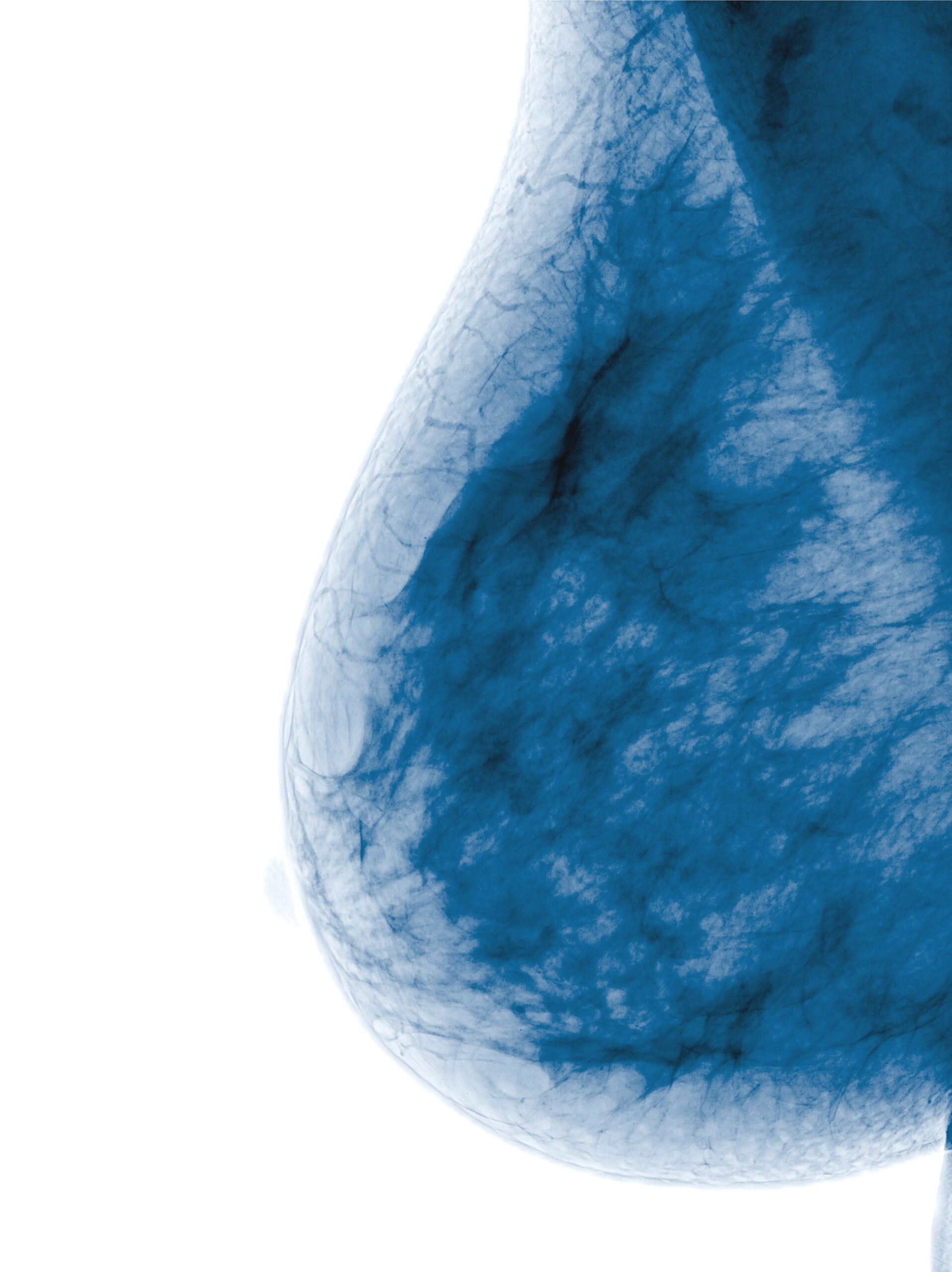 The course begins with an overview of mammography equipment going as far back as the 1960s. Essential steps to producing a quality mammogram image are reviewed as are instrumentation, mammography tubes, mammographic target materials, Bremsstrahlung interactions, photoelectric interactions, anode angles, and anode heel effect.